|
|||||
|
|
WHAT ARE THE COMPONENTS OF THE IMMUNE SYSTEM?
|
||||
|
|||||
|
|
WHAT ARE THE COMPONENTS OF THE IMMUNE SYSTEM?
|
||||
|
|
|
IMMUNE SYSTEM COMPONENTS. |
||
| PRIMARY ORGANS: (lymphocyte production and differentiation) | BONE MARROW: B LYMPHOCYTE | |
| THYMUS: T LYMPHOCYTE | ||
| SECONDARY ORGANS: (capture and processing of antigens) | SYSTEMIC: |
|
| MUCOSAS: | GROUPED:
|
|
ISOLATED:
|
||
| There are two different types of lymphoid organs depending on their function in the pig: |
THE PRIMARY LYMPHOID ORGANS are comprised of the bone marrow and the thymus. Their function is the production and regulation of the different lymphocytes. in the bone marrow, Maturation of the B lymphocytes is produced in the bone marrow, and T lymphocytes mature in the thymus.
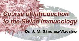
Histological section at the stroma level of the porcine bone marrow (200X) in which the different populations of hematopoietic cells can be observed.
Bone marrow: it is considered as the main lymphopoietic organ in the pig, as well as in other mammals. It is found in the inner part of every bone in the animal, especially in the long bones. There, the lymphopoietic stem cells are produced, which will give rise to the future macrophages and B and T lymphocytes. T-cells migrate from the embryionic bone marrow to the thymus cortex, were they undergo maturation.
From both an structural and a functional point of view, the porcine bone marrow is comprised of two large compartments which are related to each other:
- Stroma
- Hematopoietic
The bone marrow stroma consists of a fiber framework and reticular cells which have the role of supporting the hematopoietic part of the marrow, as well as the production of different growth and development factors for the activation of the stem cells produced in the hematopoietic part. The stroma has been defined as the hematopoietic microenvironment needed for the correct operation of the producing part of the marrow.
The hematopoietic area, in the parenchyma of the bone marrow, is formed by cell colonies or nests located in the reticular fiber net. The stem cells are called "Colony Forming Units". These cells will give rise to different cellular lines, forming the main hematopoietic organ. It is not yet known with accuracy how they begin their cell differentiation path to become one or other cell type. In addition to the bone marrow, porcine B cells are also produced, in a lesser quantity, in another lymphoid organ: mucosa-associated lymphoid tissue and spleen.
The thymus is a very important organ in the development of the immune system during the embryonic stage due to its role in the selection of self lymphocytes (associated to the SLA of each animal), as well as during the first months in the animal's life.In the young pig, it extends caudally from the digastric muscle, along the carotid arteries, at both sides of the neck, until the thoracic entry, where both thymus sides merge. It is an organ that reaches its main size between the fifth and eighth month of life, and later disappears gradually after the first year of life.
In the pig, the thymus has a lobulated structure, surrounded by a thin layer of connective tissue, that extends itself in thin septae that divide the lobules into several sub lobules which are partially separated from each other. From a histological and functional point of view, we can differentiate two zones: A cortex, essentially formed by a great accumulation of proliferating lymphocytes of different sizes (from 5 to 9 micrometers) and a scarce presence of epithelial cells, and a medulla, constituted by numerous epithelial cells that form concentric structures (called Hassall's corpuscles), and T cells in a smaller number than in the cortex.
The learning process of the lymphocytes for the acquisition of self tolerance and the capability of reacting against foreign molecules, is mainly achieved during their passage from the cortex to the medulla in the thymus.
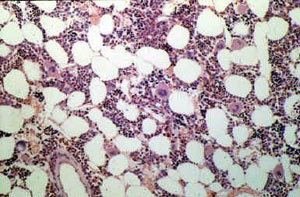
Histological section of the pig thymus (25X) in which a lobule and its sublobules can be appreciated, with the dividing septae (a), the cortical zone or cortex (b) with a great presence of lymphocytes, the medullar zone or medulla (c), formed by epithelial cells and a small number of lymphocytes.
The thymus is the first organ developed from the stem cells of the bone marrow. Once in the thymus, this cells differentiate into T lymphocytes that remain in the thymus, or migrate to other secondary lymphoid organs, forming the T- dependent zones.
|
|
|
|
|
|
|
|
|
The main function of these organs is to perform the recognition of the antigens and the initiation of the immune response. |
|
Lymph nodes: Lymph nodes are nodular formations of dense connective tissue, of a whitish color, lying along the course of lymphatic vessels, forming the ganglion chains. Their function is to capture the antigens that arrive from the lymphatic fluids and proceed to their presentation and antigenic processing by means of a collaboration between the macrophages and the lymphocytes that constitute them. The pig has a peculiar lymphatic circulation and cellular disposition, different from other mammals. |
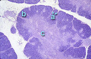
Schematic views of a pig lymphoid node, where it can be appreciated the lymphatic circulation and cellular distribution, different to that of the rest of mammals. The cortical zone, with its lymphoid follicles and its diffuse lymphoid tissue, is located in the central part of the node, being the medulla in the periphery. The lymph passes into the node through the concave surface (afferent vessel) and exits the node through the convex surface (efferent lymph vessel) |
|
From a histological and
functional point of view, two different areas can be differentiated in the
node parenchyma: cortical tissue and medullar tissue. The diffuse lymphoid tissue is considered as a T- dependent zone. It has a stroma formed by fibers, and reticular cells that constitute a three- dimensional framework where, besides T- lymphocytes, macrophages and dendritic cells can be observed, . The medullar tissue is distributed in the periphery of the node units, and around the efferent hilus. It mainly comprises fixed cellular elements, reticular fibers, and collagen. It does not present, contrary to other species, either medullar strings or sinuses. It forms a diffuse and uniform framework with less permeability that in other species. It is poor in lymphocytes, even though macrophages and dendritic cells can be observed. |
Histological section of a lymph node in the pig (25 X) in which the special distribution can be observed; the cortical zone (a) is located in the central area, with a great presence of lymphoid follicles (b) (B and T CD4+), and diffuse lymphoid tissue (c) and the medullar zone is in the periphery (d). |
|
|
|
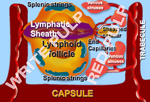
Spleen diagram. |
The porcine spleen has a framework formed by a thin capsule of muscular fibers and collagen. This supports the functional part of the spleen, consisting of: white pulp and red pulp. The white pulp is the lymphoid tissue of the spleen, always located around an artery or arteriole, forming lymphoid follicles and periarteriolar lymphatic sheaths. The lymphatic follicles consist of: lymphocytes (mainly B lymphocytes), some plasmatic cells and cells that present the antigen (macrophages, dendritic cells). The periarteiolar lymphatic sheaths are, however, formed by T lymphocytes, being some B lymphocytes in the more peripheral zones. One of the more significant characteristic of the porcine spleen is that among its T lymphocyte populations, the more numerous is the CD4+, much more than the CD8+. |
 Histological section of a porcine spleen (100 X), where the capsule (a), the white pulp (c) and the trabeculae (d) can be seen.
|
The red splenic pulp
is formed of three different structures: The splenic strings, the venous
sinuses and the sheathed capillaries. |
|
Mucosal-associated lymphoid tissue. We know as mucosal-associated lymphoid tissue the immune system along the mucosas, that have a certain independence from the systemic immune system. When along the porcine intestinal tract, it is called "gut associated lymphoid tissue", GALT. The mucosal-associated lymphoid tissue consists of lymphoid nodes that have the mission of protecting the pig mucosa from pathogenic aggressions. The mucosal-associated lymphoid tissue has a major role due to the fact that in the pig, a large number of pathogens use the mucosa as a way of entry into the animal. Around an 80% of their action mechanisms are mediated by the isotype A immunoglobulins (IgA), except in the tonsils, where the mayor synthesized immunoglobulin is the IgG, followed by the IgA. The most organized structures of the porcine GALT are: |
|
|
|
|
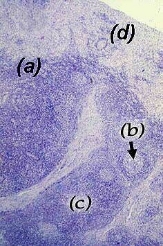
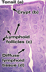
The Tonsils are part of the secondary lymphoid organs. Due to their location in the soft palate, between the respiratory and digestive tracts, they are part of the immune system of the respiratory and digestive mucosae. They are of great importance in porcine defensive mechanisms against infectious agents.
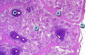
Histological section of the pig tonsil (25 X), where it can be observed: stratified epithelium (a), crypts (b), lymphoid follicles (c) (consisting mainly of B cells) surrounded by diffuse lymphoid tissue (d), chiefly formed by T cells. |
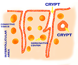
Diagram of the pig tonsils. Their inner distribution can be seen, formed by the crypts, with different appearance depending on the section, and the lymphoid follicles, with their germinal centers and the interfollicular tissue or diffuse tissue. |
|
|
They consist either of a solitary node or an aggregation of nodes and diffuse lymphoid tissue. They are located in the lamina propia, closely associated with the epithelium, which depending on its situation, can be: stratified or pseudostratified in the nasopharynx. The tonsil's surface can be smooth or with deep invaginations, as is the case of the pig epiglottis tonsil. This allows a large quantity of lymphoid cells to concentrate in the invaginations.
|
||
|
The lymphoid structure of the tonsils is similar to that of the lymphoid follicles. There are germinal centers that are confined and compact structures. They are formed by a large number of B cells which are included in the diffuse lymphoid tissue, mainly formed by T lymphocytes. All this is closely associated with the epithelium. The antibodies mainly produced in the tonsils are IgG, followed by IgA. |
||
|
Peyer's
patches. They
are lymphoid tissue aggregates, non encapsulated, and located
in the gut submucosa. In the pig they can be separated by their size and localization in two different types:
Ileocecum Peyer's patches: terminal ileum. |
||
|
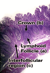
Histological cut of a Peyer's patch (25 X). The lymphoid follicles can be appreciated, with their germinative centers (a), the crown (b) and the interfollicular region (c).
|
From a histological and functional point of view, we can differentiate four zones in the Peyer's platelets:
|
|
|
Jejune Peyer's patches: They are small size patches (from 25 to 35), distributed along the jejune and the proximal portion of the ileum. They consist of B and T lymphocytes and remain during the pig's lifetime. The ileumcecum Peyer's patches: They are of a large size and are located in the terminal portion of the ileum. They diminish during the first year of life. Their cellular composition also differs from the jejune patches; in the ileumcecum Peyer's patches there are ten times more B cells than T cells. |
||
|
It is important to note that along the porcine intestinal tract, especially in the stomach and large intestine, and in both the mucosa and in the submucosa, isolated lymphoid follicles can be found. They are covered by an epithelium which is similar to the one that covers the Peyer's patches, and their function also seems to be similar. |
|
|||||