|
|
PORCINE IMMUNOGLOBULINS |
|
|
|
PORCINE IMMUNOGLOBULINS |
|
The immunoglobulins are a family of proteins produced by B lymphocytes when they are activated by an antigen (plasma cells)
They can be found in the serum and tissue fluids of every mammal, both as a secretion (ANTIBODIES) or linked to the B lymphocyte membrane (BcR RECEPTOR). They mediate in the humoral response. Four different types of immunoglobulins have been described in the pig. They are called: IgM, IgG, IgA e IgE. |
||
| HOW ARE IMMUNOGLOBULINS PRODUCED? | ||
|
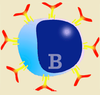
Immunoglobulins are produced after the stimulation of a B lymphocyte by antigens (both by T independent and T dependent antigens) and by the transformation of this B lymphocyte in a plasma cell. Plasma cells do not divide, neither do they change their isotope nor express SLA II or surface immunoglobulins. Thus, they can not interact with any type of antigen. They are factory cells for the production of immunoglobulins. |
Diagram of a B lymphocyte and its activation to plasma cell. All immunoglobulins produced by each plasma cell clone are specific against the epitope that induced the immune response. There will be as many antibody producing clones as inducing epitopes. When an antigen (consisting of several epitopes) induces an immune response, there will be different plasma cell clones segregating specific antibodies against each epitope. |
|
|
Due to the great structural complexity of most antigens, when a humoral response is produced against any of them, a very large number of different antibodies is induced, targeted to the different epitopes of the inducing antigen. Thus the immune response is polyclonal, because there are thousands of stimulated clones that secrete different antibodies.
|
||
| WHAT IS THE STRUCTIRE OF THE IMMUNOGLOBULIN LIKE? |
||
|
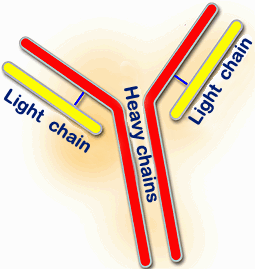
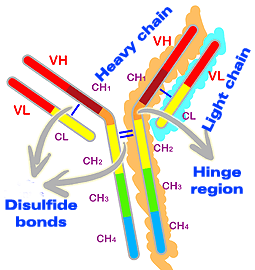
Porcine immunoglobulins are, just as in other species, glycoproteins consisting mainly of four polypeptide chains. Two of them
of approximately 55-77 kilodalton (KDa). They are termed HEAVY or H chains.
The other two of 23 to 26 KDa are termed LIGHT or L
chains. Both types of chains have an identical structure. Heavy chains are covalently linked to
each other by disulfide bonds and the heavy chain is linked to the light one also by disulfide
bonds. Each chain has a constant and a variable region. |
|
|
|
|
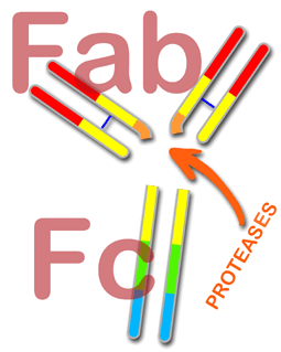
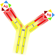
When the immunoglobulin is treated with proteolytic enzymes (proteases), such as pepsin or papain, it is broken into two fragments known as Fab (for antigen binding Fragment) and Fc (for Crystalizable Fragment). The immunoglobulin specifity is determined by the Fab fragment, as well as its capability to react with the antigen. The effector role is performed by the Fc fragment (complement activation, cell receptors, etc...) |
|
|
Both heavy and light chains consist of some conserved protein structures known as Immunoglobulin Domains. These domains correspond to approximately 110 amino acids. Light chains have two domains, one of them variable (VL) and the other constant (CL). Heavy chains have one variable part (VH) and three (IgG and IgA) or four (IgM and IgE) constant (CH1, CH2, CH3 y CH4). These domains are identical between the two light chains and between the two heavy chains. Heavy chains have an additional region, that does not form part of the domains. It is known as the hinge region. This hinge region is located between the CH1 and CH2 domains and givess flexibility to the immunoglobulin. When the amino acids of the hinge region are analyzed, high amounts of proline can be found. This gives flexibility, but also susceptibility to being attacked by protease-enzymes. This allows breaking into Fab and Fc fragments.
|
The variable domains (VL and VH) contain
the antigen-binding activity, and are therefore responsible for the immunoglobulin specifity.
Furthermore, the constant domains allow the diversity of the five heavy
chains isotypes (m,g,e,a,d) that will determine the immunoglobulin type (IgM,
IgG, IgE, IgA and IgD) as well as the two
light chains: kappa (K) and lambda (l). They are also responsible of the effector
functions of the immunoglobulins (complement activation, cell receptors, etc...) |
|
|
|
The variability observed in the variable regions of both chains (L and H) is located in three segments of about 10 amino acids that are designated hypervariable regions. They are also known as: CDR1, CDR2 y CDR3 (Complementary Determining Regions). These fragments form the binding site for antigens i.e.; the antigen-binding site. Therefore, each immunoglobulin molecule has two antigen-binding sites. |
|
|
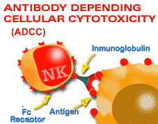
In addition, carbohydrates play an important role in the immunoglobulin structure, especially in the constant region of heavy chains (mainly in CH2 zone) and in the hinge region. However, carbohydrates make up only about 15% of the heavy chains. The precise role of carbohydrates is not well known, but they may be related to catabolism mechanisms and to some of the immunoglobulin functions. It has been shown that deglycosilated immunoglobulins lose or lower their ability for binding to cell receptors, of inducing ADCC, and of complement activation. |
|
|
|
© Copyright. 2001. José Manuel
Sánchez-Vizcaíno Rodríguez. All rights reserved.
Dep. Legal: B-32.422-01. ISBN: 84-699-5917-4