|
|
PORCINE LYMPHOCYTE CHARACTERISTICS. |
|
|
|
PORCINE LYMPHOCYTE CHARACTERISTICS. |
|
|
||||||||||
|
SELF RECOGNITION, SPECIFITY AND IMMUNOLOGICAL MEMORY. |
||||||||||
|
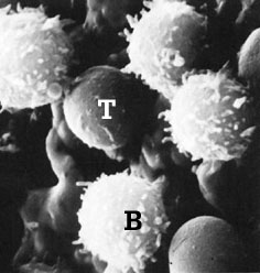
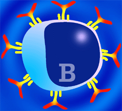
When using an ordinary microscope, no differences can be found (from a morphological point of view) between T and B lymphocyte. Both lymphocyte types are cells from 7 to 9 micrometers, with a voluminous nucleus and a little cytoplasm. Using scanning electron microscopy, remarkable differences can be observed. T lymphocytes present a smooth and even surface while B lymphocytes have multiple projections, (these are the surface immunoglobulins). |
|
|||||||||
|
T and B lymphocytes by scanning
electron microscopy |
||||||||||
|
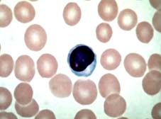
Giemsa stained lymphocyte, observed by ordinary microscopy |
In the last decades, different surface markers to differentiate among lymphocyte populations have been designed. However, the development of monoclonal antibodies (MAb) against the different lymphocyte porcine cells have permitted the differentiation of these populations, as well as their inclusion in different groups depending on their membrane antigens and functions. |
|||||||||
| MAb have
also allowed the classification of a third lymphoid population, known before as null cells due to their absence of
traditional T and B lymphocyte markers (rosette
formation with sheep erythrocytes and superficial immunoglobulins respectively) These
null
cells, which could compose around 9 to 19% of porcine circulating lymphocytes, are in fact a
T lymphocyte subpopulation (T lymphocytes g-d)
|
||||||||||
|
|
|||||||||
|
|
||||||||||
|
B lymphocytes of the pig are produced in the bone marrow, in a
proportion of two hundred to four hundred million a day. This shows the huge
capacity of response of
the immune system. You must remember that each lymphocyte secretes a specific
antibody (1 cell = 1 antibody type). In the peripheral blood, B lymphocytes
make up 8-18% of total lymphocytes. The lymphocyte membrane has a large number of molecules, many of them having
being studied thanks to MAb. Among them it is important to underline the importance of B Cell Receptor
complex or BcR complex. |
B lymphocyte |
|||||||||
|
BcR consist of several chains. Some of them are variable (they are immunoglobulins), in which each lymphocyte presents different variations depending on the immunoglobulin type (mainly IgM and IgG, but also IgA or IgE) or on the antigen type. |
||||||||||
|
|
The remaining two chains are constant (consisting of two chains, a and b), common to every B lymphocyte. The function of the variable chains, which in fact are immunoglobulins, is to interact with the specific antigen, while constant chains transmit the signal to the cell interior in order to initiate the antibody production. B lymphocytes are thus characterized by having immunoglobulins on their surface, mainly IgM and IgG. This allows their interaction with the antigen in its native form (T lymphocytes can not react with an antigen in its native form) (Chapter 3). The majority of the antigens that react with B lymphocytes are proteins, although they can also interact with polysaccharides. The initiation of the antigen reaction is always through the membrane immunoglobulins, (BcR signal), but antibody production needs, in most cases, the collaboration of CD4+ lymphocytes (chapter 3). The antigens that need this collaboration are known as T- dependant antigens, and the majority of antigens belongs to this group. Other antigens, such as some bacteria polysaccharides or lypo polysaccharides, can produce antibodies without the collaboration of T lymphocytes. These are known as T- independent antigens. |
|||||||||
|
|
||||||||||
|
||||||||||
|
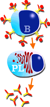
Any stimulated B lymphocyte, either by a T- dependent antigen or by a T- independent antigen, generates a plasma cell clone, which will produce and secrete a large quantity of antibodies. These antibodies are specific for the epitope that induced the immune response. This response, mediated only by antibodies, is known as humoral response. The stimulated B lymphocyte, or plasma cell, has a morphology different from the original B lymphocyte (large nucleus and small cytosol) and more similar to a "factory- cell", with a small nucleus and a large cytosol. These cells are mainly located in lymph nodes, spleen red pulp, bone marrow and gut and respiratory mucosa. Their "in vivo" mean life is short: 2 or 3 days. It is even shorter "In vitro, only a few hours. |
|
|||||||||
|
B lymphocyte stimulation, transformation in plasma cell, and antibody secretion. |
||||||||||
 |
Porcine T lymphocytes are produced in the spleen, and in smaller quantities in the T-dependent zones of secondary lymphoid organs. They differ from B lymphocytes in the sense that they do not present surface immunoglobulins, but form rosettes with sheep erythrocytes. T- lymphocytes play a main role in the immune response. In the first case, in the antigen-presenting mechanisms for B lymphocytes to produce antibodies, and in the second case, they are responsible for cellular immunity (immune response mediated by cells, not by antibodies) (Chapter 7). |
|||||||||
|
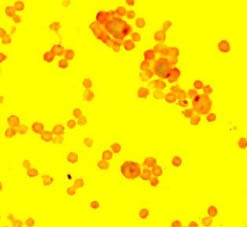
Rosettes formation by T lymphocytes with sheep erythrocytes. |
||||||||||
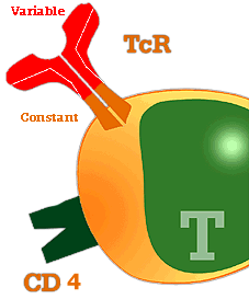
There are two more types of antigen specific receptors in the T lymphocytes membrane apart from SLA. These are the T cell receptors or TcR: |
||||||||||
|
Receptor a-b (TcR a-b). They comprised 40 to 60% of the lymphocytes present in peripheral blood. T
LYMPHOCYTES a b |
||||||||||
|
Just as in the case of B lymphocytes, the production of MAb against porcine lymphocytes, has made it possible to differentiate several T cell subpopulations. They have been grouped in clusters of differentiation (CD) depending on their antigenic and functional differences. |
|
|||||||||
| TcR complex of porcine lymphocytes. It consists of a variable part that reacts with the antigen, and a constant part. | ||||||||||
|
Thanks to these markers, it has been confirmed that pigs present some differences in respect to other animal species and humans.. Thus, the pig has a lymphocyte subpopulation which is double positive to CD4 and CD8 (CD4+CD8+). The percentage of this population increases with age. In this way, when the animal is one week old, this percentage is smaller than 2% of the total lymphocytes. When it is 3 years old, the percentage is 30%. The role of these double positive lymphocytes was thought to be related to memory cells. However, now it seems they are related to the helper activity of T lymphocytes during primary infections. It seems that there are two different populations of helper T cells, one being CD4+CD8- and the other CD4+CD8+. Both populations act as helpers during the primary response (at least, they do so "in vitro"). During the secondary response only CD4+CD8+ seem to have a role.It is also known that these double positive populations not only increase with age, but they are also located in large proportion in the tonsils (50%) and lymph nodes (30%) in adults. In regard to cytotoxic T lymphocytes, two different subpopulations of DC4-CD8+ can be found depending on the expression of CD6. Thus, lymphocytes expressing CD6 antigen are related to spontaneous cytotoxicity, while CD6+ SLA I are related to virus-infected cells cytotoxicity. |
|
|||||||||
| Finally, there is another special characteristic of porcine T lymphocytes: they express SLA II in their membranes. This is different in humans or mice, whose lymphocytes do not present class II histocompatibility antigen when they are not activated. | ||||||||||
|