|
|
THE OTHER CELLS |
|
|
|
THE OTHER CELLS |
|
This group of cells is made up of:
|
||||||||||||
|
||||||||||||
| THE ROLE OF PHAGOCYTIC CELLS. | ||||||||||||
|
|
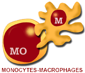
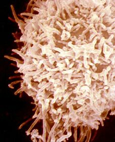
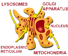
Monocytes-macrophages. They are large cells, with a diameter of around 15 mm, with a large cytoplasm and a single nucleus that can be spherical, ovoid or lobulated. A very developed Golgi apparatus can be observed in their cytoplasm, as well as the rough surface of the endoplasmic reticulum, mitochondria, and a large number of lysosomes, which contain hydrolytic enzymes. This means that they have a great capability of producing and secreting proteins. They derive from the myeloid lineage. First, they
undergo transformation into promonocytes in the bone marrow; then
into monocytes, in periphery blood and finally into macrophages in the different organs.
|
|||||||||||
|
Macrophage diagram. |
||||||||||||
|
|
||||||||||||
|
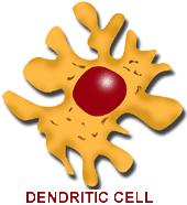
Monocytes-macrophages, together with dendritic cells and B lymphocytes, are the only porcine cells that express in their surface histocompatibility antigens SLA II. Thus, they are the only cells able to present antigens to helper T lymphocytes CD4+. This cellular family is known as Antigen- Presenting Cells (APC). |
||||||||||||
|
Monoclonal
antibodies are used nowadays to study and differentiate monocytes-macrophages. In the
last Symposium about porcine immune system differentiation, in 1998, three monoclonal antibodies were
defined for the study of porcine macrophages. They were called SWC (for Swine
Workshop Cluster), plus a number. Thus,
the most used Mab at the moment are: SWC1 and SWC9, which mainly differentiate monocytes
and macrophages, and SWC 3 that react both with monocytes and with differentiated
macrophages and granulocytes.
|
||||||||||||
|
These markers have recently allowed us to come to know that monocytes are SCW1+SWC9-. When transformed to macrophages however, they become SWC1-SWC9+. Different studies in peripheral blood monocytes can be done by means of flow cytometry, while for tissue studies, histochemical techniques are performed (How are cells studied?) |
Macrophages Besides presenting a characteristic morphology depending on the organ where they are located, different types of activity, depending on their maturation level, their activation, and their specific localization. |
|||||||||||
|
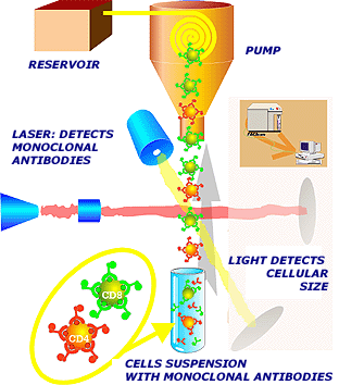
Suspended cells, with monoclonal antibodies added , pass through a very thin tube, forming a single line of cells. They are analyzed both by laser and light beams. First, the laser beam identifies labeled antibodies, and the light beam, measures cell size. |
||||||||||||
Among these activities, we can underline: |
||||||||||||
|
||||||||||||
|
Macrophages have the function of phagocytosis and lysis of microorganisms and infected or tumor cells, acting both directly (natural or innate immunity) or through their Fc receptors for immunoglobulins or for the complement. As antigen presenting cells, they participate in the acquired immunity, binding and processing antigens in order to present them to T lymphocytes. Finally, macrophages produce a large number of cytokines: interleukines 1, 2, 6, 12, interferon a and b, tumor necrosis factor-a (TNF-a) (Chapter 6). They also produce the complement elements as well as several enzymes (Chapter 7). |
||||||||||||
|
|
|
|||||||||||
|
|
||||||||||||
|
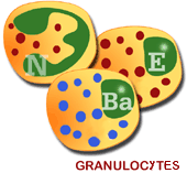
Neutrophil or Polyimorphonuclear |
||||||||||||
Four different stages can be
defined in the phagocytosis, even though it is a continuous process. These stages
are:
A Macrophage
phagocytizing two bacteria |
|
|||||||||||
|
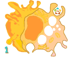
Phagocytosis activation occurs by the liberation of chemotactic substances (chemotaxis). Tthe complement is activated when platelet factors are liberated or by means of other substances. Once the macrophage encounters a foreign particle, it binds to it neutralizing its negative charges (both are repelled). This phenomenon is eased when the foreign particle is attached to immunoglobulins or the C3b fraction of the complement (Adherence). The ingestion occurs by means of pseudopoda, which enclose the foreign particle, incorporating it to the cytoplasm and forming a vacuole or phagosome (Ingestion). Finally, the particle is digested by lysosomal enzymes (Destruction). |
||||||||||||
|
The second most important granulocytes are the Eosinophils. They are so named because they incorporate acid histological stains, such as eosin. Eosinophils enter the spleen from the bone marrow when they are not completely mature, and then undergo maturation in this organ. Later, they pass to blood and tissues. Their half live is very short, less than an hour. Their main role is also phagocytic, even though their granules do not contain lisozime.They do have large quantities of acid phosphatase and peroxidase. They are very effective against parasites. In summary, even though granulocytes do not react in a specific way with antigens (recognition of antigens depends on innate reactions usually due to the receptors of the own pathogen or to complement activation) they play a main role in phagocytic and inflammatory processes, especially when these have an acute character. Their membrane receptors allow them to phagocyte opsonized particles.Thanks to their receptors for the Fc fraction of the immunoglobulins, they can act as effector cells in antibody depending cellular cytotoxicity (ADCC) |
|
|||||||||||
|
||||||||||||
| Lastly, the least abundant granulocytes are
the basophils, which are so named due to being stained by basic stains, such as
hematoxiline. Their activity is related to the liberation of vasoactive amines
such as histamine and serotonin.They intervene in acute inflammations, acting as a warning signal for the immune system.
|
||||||||||||
|
© Copyright. 2001. José Manuel
Sánchez-Vizcaíno Rodríguez. All rights reserved.
Dep. Legal: B-32.422-01. ISBN: 84-699-5917-4