|
|
THE MUCOSA ROLE
|
|
||||
|
|
THE MUCOSA ROLE
|
|
The mucosa play a very important role in the immunological defense of a pig. This is because a large number of pathogenic agents are able to use the mucosa as an entry way.
The lymphoid tissue associated
to the mucosa is part of the immune
system, although it works somewhat independently of the systemic system. It is the one that protects the pig mucosa from the attack of
pathogenic agents, both in the primary and secondary response. It consists of lymph nodes that, depending on their situation, are called: GALT
and BALT |
||||||
|
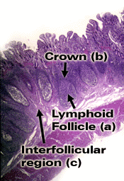
GALT is an acronym for: "Gut Associated Lymphoid Tissues". The GALT consist of the lymphoid tissue located in the epithelial surfaces of the gastrointestinal tract (lymph nodes, Peyer´s patches, isolated lymph follicles). (Secondary lymphoid organs) BALT comes from, "Broncus Associated Lymphoid Tissues", and consist of the lymphoid tissue of the respiratory mucosa, from the nose to the lungs (tonsils, lymph nodes, lymph follicles). |
|
|||||
|
There is one peculiarity in the porcine BALT; there are large quantities of very active intravascular macrophages in the lungs. This does not happen in the human and murine species. |
||||||
|
MUCOSA IMMUNITY |
||||||
|
The large amount of mucosal-associated lymphoid tissue in the pig shows the importance of this tissue in the defense against infections This lymphoid tissue is strategically distributed in the following locations:
The initiation or inducting
areas have similar elements to those of the systemic immune system for
trapping antigens and beginning the immune response. With the sole exception
of the M cells, which are epithelial cells specialized in antigen transportation, the rest of the
cellular components (antigen presenting cells, T and B lymphocytes) act in a similar way to the
systemic immune system. They are located in the tonsils, Peyer´s platelets and diffuse lymphoid
tissue. In summary, antigen binding, transportation, processing and T and B presentation take
place in GALT and BALT areas (see secondary lymphoid organs of the
pig) |
||||||
|
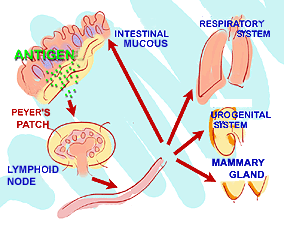
This mechanism enables a generalized response even though the antigenic stimulation has been in a local level. This immune response is known as the:generalized secreting response. |
|
|||||
|
Stimulation of mucosal-associated lymphoid tissue, BALT or GALT. This mechanism allows a generalized response even when the antigenic stimulation has been a local process. |
||||||
|
|
||||||
|
IgA immunoglobulin |
Most of the immune cells present in the effector areas are T lymphocytes (mainly CD8, around 77%, and DC4 g-d) which are located among epithelial cells or beneath them, in the lamina propria. Some B lymphocytes are also present and they can react with the antigen. Plasma cells, secreting mainly IgA immunoglobulin, are situated in lymph nodes and in diffuse lymphatic tissue of gastrointestinal and respiratory walls. These cells play a major role in the mucosa immune response, secreting about 80% of the IgA produced, with the exception of the tonsils, where IgG is the predominant immunoglobulin, followed by IgA. |
|||||
|
|
||||||
|
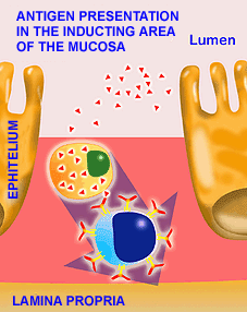
Antigen transportation to the inductor areas (Peyer´s platelets and lymphoid follicles) is mainly done by what are called M-cells. M-cells are epithelium cells specialized in antigen transportation. They do not act enzimatically against antigens. M-cells trap antigens in the gastrointestinal lumen and carry them to epithelial lymphocytes or enter through the inter-cellular gap to the extracellular fluid, where they present the antigen to APC (macrophages, dendritic cells and B lymphocytes) of the sub-epithelial area or lamina propria. The activation mechanisms in the lamina propria follow a similar pattern to the one for the cellular cooperation described above. |
|
|||||
|
|
||||||
|
|
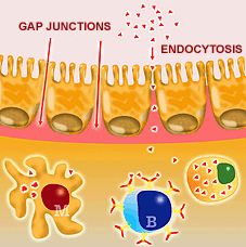
Antigen presentation can also occur in the effector areas, although the entry mechanism is usually different to that of the inducting areas. Antigens can enter the effector area by endocytosis or through gap junctions. Antigen binding and presentation are performed by macrophages, M-cells or B lymphocytes, and the subsequent stages follow the same mechanisms described above. The immune response induced in the mucosa usually needs a larger quantity of antigen, and sometimes also a greater number of immunizations, than that of the systemic system. This is especially true in the case of oral immunizations. |
|||||
|
Antigen in the effector area, where it enters by endocytosis mechanisms or through the gap junctions. |
||||||
|
This is due to the antigens suffering a series of enzymatic alterations and degradations when they enter the organism by this mechanism. This mechanism is good for the animal immune defense but must be taken into account when preparing oral vaccines. There are however, several strategies used to produce a good oral immune response (chapter 8). Nevertheless, the induction of immunity in the respiratory tract is generally easier by an oral immunization than by producing immunity in the gastrointestinal mucosa by a nasal immunization. |
||||||
|
|
IgA (chapter
4) plays a major role in the mucosa immune response. Its dimeric or
tetrameric structure allows 4 to 8 immunoglobulin binding sites. This makes it tremendously effective against different bacterial antigens by means of ADCC
reactions;
IgA is not a bactericide. It does however, have the capability of neutralizing several viruses,
even inside epithelial cells. In fact, it is the only immunoglobulin able to work in the cell
interior. Nevertheless, the main activity of the IgA in the mucosal defense is to avoid the attachment of
bacteria and viruses to the epithelium surface. Thus, the IgA can have its activity in three different
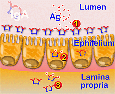
ways: firstly, it can bind the antigen in the gastrointestinal lumen,
preventing antigen attachment to the epithelial surface; second, it can act inside
enterocytes, and finally, in the extracellular fluid. |
|||||
|
DIAGRAM OF IgA ACTIVITY MECHANISMS: IgA prevents the attachment of bacteria or viruses to epithelial cells in the gastrointestinal lumen (1); virus neutralization inside enterocytes (2); in extracellular fluids. |
||||||
|
Another example of the importance of this immunoglobulin in the defense mechanisms of the porcine species is the fact that 85% of immunoglobulin containing cells of the pig intestine lamina propria have IgA. |
||||||
|
||||||